Optomap retinal exam
Mae'r cynnwys hwn ar gael yn Saesneg yn unig.
The Optomap retinal exam gives us a wide view to look at the health of your retina, producing an image that is as unique as your fingerprint.
The retina is the part of your eye that captures the image of what you are looking at, similar to film in a camera. Optomap retinal imaging uses a camera that gives optometrists a 200 degree view of the back of the patient's eye (compared to an approximately 60 degree view). This gives a much better picture of the retina than conventional cameras which most opticians with an imaging system still use.
Optomap retinal imaging offers a number of important benefits, including:
- a wide field of view of the retina which cannot be achieved with conventional cameras
- a permanent record of your retina for future comparison
- early detection of eye diseases and disorders
- no need for dilation drops
- a non-invasive, stress-free and comfortable process
- it's suitable for all ages (including children)
- you can have Optomap taken even if you have cataracts, glaucoma, small pupils or don't like bright lights.
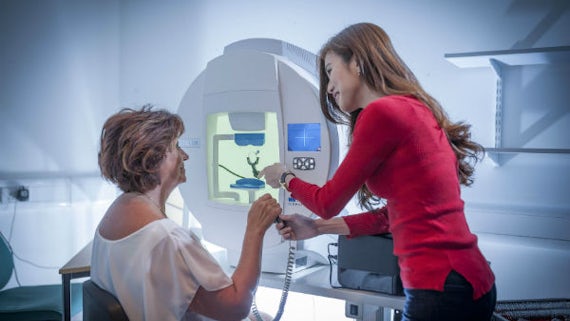
To be examined, you will look through a scanning instrument at a green spot surrounded by a red outer circle. When you see that the circle has become red, an Optomap photograph is taken. It takes less than a second to take an Optomap photograph of the back of the eye. The optometrist then analyses the photograph to check for any eye problems.
An Optomap exam is £20.
Cysylltwch â ni ar +44 (0)29 2087 4357 neu anfonwch e-bost atom i drefnu apwyntiad.