Magnetic Imaging
Understanding magnetic materials and nanostructures is frequently aided by utilising a range of imaging techniques and modalities.
In addition to using many standard magnetic imaging techniques in the labs at Cardiff we also make use of synchrotron radiation and other central facilities worldwide. We are also actively developing new scanning probe microscope (SPM) techniques to apply to magnetic imaging as a function of temperature and other physical parameters.
Activities
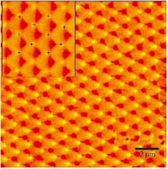
Standard magnetic imaging techniques available at Cardiff are both Magnetic Force Microscopy (MFM) and Magneto Optic Kerr Effect (MOKE) which have both been used to understand a range of material systems. These studies have included artificial spin ice structures, magnetic domain walls in nanowires being developed for prototype next generation data storage, and magnetic sensors.
To achieve the highest resolution images with magnetic contrast Cardiff researchers are regular users of large scale facilities including Diamond Light Source in Oxfordshire, UK and the Advanced Light Source in Berkeley, USA.
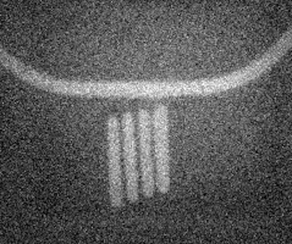
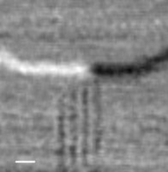
Synchrotron techniques allow the use of X-ray Magnetic Circular Dichroism (XMCD), which can allow the magnetic domain structure of samples to be measured on nanometer lengthscales. Additionally due to the fact that we can normally tune the incident x-ray energy these techniques would allow an element specific measurement of the magnetism in materials containing a variety of chemical species.
Cyhoeddiadau
- Zeissler, K. et al., 2013. The non-random walk of chiral magnetic charge carriers in artificial spin ice. Scientific Reports 3 1252. (10.1038/srep01252)
- Ladak, S. et al. 2010. Direct observation of magnetic monopole defects in an artificial spin-ice system [Letter]. Nature Physics 6 (5), pp.359-363. (10.1038/nphys1628)