Bioimaging Research

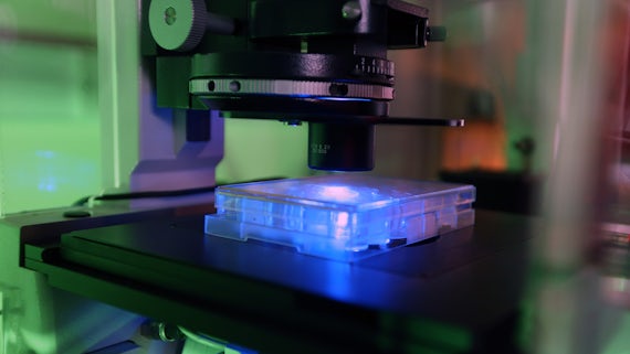
The Bioimaging Research Hub provides state of the art technology and expertise in light microscopy, from experimental concept to completion and is linked with the Biophotonics Laboratory in the School of Biosciences as well as other equipment spokes.
The Hub works in partnership with researchers across Cardiff University and GW4 member institutions. We also provide innovative imaging solutions across biological, chemical and material science to the industrial sector.
Full technical support, training and advice on the use of all of our imaging equipment is available to users on a chargeable basis.
Our services
We offer a number of histological processing and slide preparation services, including:
- Paraffin wax processing and embedding
- Wax microtomy and slide preparation
- Histological staining - routinely Haematoxylin and Eosin (H&E) but contact the Bioimaging Research Hub at bioimaginghub@cardiff.ac.uk to discuss other staining methods.
All samples submitted for histology must be accompanied by a Histology Request Form.
People wishing to use the service should email the Bioimaging Research Hub at bioimaginghub@cardiff.ac.uk for a quote.
The Hub is home to a Zeiss LSM880 Airyscan upright confocal microscope and a Zeiss Celldiscoverer7 automated imaging system (see below section), both equipped with advanced software for processing and analysis of 3D/4D microscopy datasets. The LSM880 Airyscan confocal microscope is suitable for:
- Confocal 3D/4D visualisation of tissue sections, live cells/tissues, and non-biological materials
- Super-resolution imaging
- Multi-channel fluorescence, reflectance & transmitted light modalities
- Tile scanning & stitching
- Fluorochrome co-localisation analysis
- Photomanipulation via FRAP, FRET etc
- Fluorescence lifetime imaging (FLIM) and fluorescence correlation spectroscopy (FCS)
A state-of-the-art Zeiss Celldiscoverer 7 imaging system allows both high throughput and multi-dimensional super-resolved imaging of biological samples from live cells to small model organisms. The system has full environmental control with pipetting port for on axis sample delivery. This equipment is capable of:
- Intelligent multi-channel, multi-format imaging (i.e., supports slides, chamber slides, dishes, plates etc)
- Automated high-throughput cell screening applications
- Multi-channel imaging with fluorescence & transmitted light modalities
- Confocal 3D/4D visualisation of live cells/tissues and tissue sections
- Super-resolution imaging
- Tile scanning & stitching
- Photomanipulation via FRAP, FRET etc
- Deconvolution and machine learning
A live cell imaging spoke of the Bioimaging Research Hub houses a spinning disc confocal system for fast, time-lapse live cell imaging applications. This system enables high speed, multi-position (x,y,z), multi-colour fluorescence and transmitted light image acquisition. Support systems for live cell imaging (i.e. gas and incubation) are also available within the facility.
A state of the art Zeiss Lightsheet Z.1 system with environmental control allows multi-view, multi-colour imaging of thick fluorescent samples with high spatio-temporal resolution.
We have a range of advanced Olympus BX and Leica DM series microscopes with digital image capture, which can be used for the following:
- Brightfield microscopy
- Darkfield microscopy
- Phase contrast microscopy
- Nomarski differential interference contrast (DIC) microscopy
- Polarising microscopy
- Epifluorescence microscopy
Our Objective Imaging Surveyor slide scanning system enables high resolution scanning and digitisation of whole tissue sections for virtual histology and pathology.
An Olympus VS200 slide scanner, located at the European Cancer Stem Cell Research Institute, facilitates high throughput, fully automated whole slide scanning of histological sections via a range of image modalities, including epifluorescence.
We offer transmitted light and epifluorescence imaging of very small samples, including whole-mounts, culture plates, gels, engineered constructs and tissue sections, as well as general purpose photography.
Our Biospace Lab PhotonIMAGER Optima has full spectrum imaging capability, from blue to near-infrared, in bioluminescence and fluorescence of living tissues.
A Thermo Fisher NanoDrop 3300 fluorospectrometer allows quantitation of picogram quantities of DNA, RNA and protein in microvolume solutions.
A Zeiss PALM MicroBeam laser microdissector, located at the European Cancer Stem Cell Research Institute, facilitates isolation of DNA, RNA and protein from histological sections (paraffin wax or cryo) and live cells.
3D printer:
- Ultimaker 3 Extended which is a dual extrusion 3D printer capable of generating high quality two colour 3D models up to 30cm tall from volume datasets.
This printer accept .stl, .obj, .dxf and .3mf 3D printer file formats.

Almost all the work in my group requires optical imaging and the service provided by the Bioimaging Research Hub is the most reliable and productive I have encountered over my 50 years at Cardiff University, or indeed of those at other establishments world wide. Collaboration with Dr Tony Hayes almost invariably results in publication of images of the highest quality.
Contact us
For more information on our Bioimaging services, please contact:

Professor Peter Watson
Director for Postgraduate Education, Professor, Academic lead of imaging facilities, Postgraduate Research Teaching Co-ordinator
Find out more about our Bioimaging Research Hub, including available equipment, services, and booking information.


