Creating filters for the medical images of the future
7 February 2024
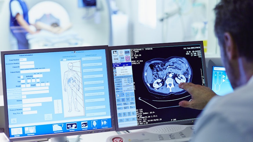
A suite of filters which can be applied to medical images to help healthcare professionals with analysis and diagnosis has been developed by an international team of researchers.
Operating in a similar way to those used on smart phones to enhance photography, the filters highlight different textures to help clinicians identify lesions or blood vessels in 3D medical images such as breast scans.
Part of the Image Biomarker Standardisation Initiative (IBSI), the work addresses a lack of standardisation in the way filters are currently applied.
Led by Cardiff University and the National Center for Tumor Diseases in Germany, the team developed a set of standard guidance and reference values for applying common filters to medical imaging.
Their study, published in Radiology by the Radiological Society of North America (RSNA), calls for a uniform approach that enhances the reliability and validity of data extracted from medical images.
The authors say this approach will benefit medical software developers and, in time, patients.
Co-lead author Dr Philip Whybra, a former research associate at Cardiff University’s School of Engineering, said: “Our research is on reproducible radiomics. These are techniques which can be used to extract measurements or biomarkers from medical imaging.
“We use these biomarkers in diagnostic and predictive models to determine the stage of a disease or to predict how a disease will respond to treatment.
“The problem right now is that different tools might produce inconsistent or even contradictory results, even when examining the same image. This can lead to potential misinterpretations and inaccuracies in medical models using imaging biomarkers.
“In our study, we’ve set about standardising the use of convolutional filters on medical images as part of a radiomics workflow.”
Part of the IBSI’s efforts to standardise image processing software, the team’s work means the application of filters to medical imaging can now be tested and verified.
This will make it easier to successfully validate artificial intelligence (AI) tools that use image filters, paving the way for their introduction in clinical practice in the future, the authors claim.
Co-lead author Dr Alexander Zwanenburg, a postdoctoral researcher at the National Center for Tumor Diseases, added: “Understandably, there is major interest in the application of AI tools in healthcare because of their potential for making processes more efficient and improving patient treatment.
“AI tools that analyse medical imaging require software to process the images and extract information from them. Our study tackles an ongoing challenge which is that available image processing software packages, created by researchers and companies around the world, often do not yield the same results.
“Due to this lack of reproducibility, many medical imaging-based AI tools cannot be validated – that is, checked if they work as advertised in new settings. Failure to be validated precludes the use of these tools in patient care.
“Now, with this hurdle removed, I hope that we have contributed to bringing useful AI tools into the clinical setting for the benefit of patients.”
Cardiff University’s contributions to the research came from the Life Imaging and Data Analytics (LIDA) Facility, led by Professor Emiliano Spezi.
LIDA focuses on advanced medical image processing, radiomics techniques and advanced computer modelling to optimise and personalise treatment delivery.
Professor Spezi, Professor of Healthcare Engineering at Cardiff University’s School of Engineering, said: “I am very proud of the contribution that LIDA has made to the IBSI.
“Research in medical image analysis is critically important to improve diagnostic accuracy, early detection, treatment planning and the healthcare system.
“This new publication from the IBSI sets another milestone towards standardisation and interoperability of non-invasive imaging biomarkers.”
Share this story

The School has world leading research, strong links with industry, and a friendly and supportive teaching environment make us one of the leading engineering schools in the UK.


