The beauty of your brain and how to see it
“Your existence, your personality, everything you do, everything you think, everything you’ve done — it’s all there in that few litres of stuff.”
In a sun-soaked corner of Cardiff University’s Brain Research Imaging Centre, a selection of sci-fi film posters hang on the wall of Professor Derek Jones’s office.
Jones is the Centre’s director and a leading expert in brain imaging.
“There’s something seductive about the brain,” he says. “You wouldn’t have The Liver That Wouldn’t Die or The Creature With The Atom Pancreas. The brain seems to feature in horror stories probably because it’s an unknown—there’s something mysterious about it.”
One poster, depicting 1981 film Scanners, is particularly eye-catching:
'10 seconds, the pain begins. 15 seconds, you can’t breathe. 20 seconds, you explode. SCANNERS… Their thoughts can kill!'
Jones jokes about moving it downstairs to welcome volunteers, but he's keen to get across that the reality of an MRI scan is more relaxing.


Into the scanner
Volunteers are asked to change into a set of custom embroidered purple scrubs to remove the chance of anything metallic following them into the scanner.
“The magnets are three or seven times stronger than the kind of magnet you get in a junkyard that lifts cars up,” explains Jones. “If it can lift a car, you can imagine how much it would pull on a coin in your pocket.”
They lie in the scanner and are asked to keep still.
A blanket gives extra comfort and earplugs protect the ears from the loud rhythmic pulses and bloopings of the scanner. Finally, an antenna-like receiver coil is slid over the volunteer’s head.
“I can’t stay awake in the scanner,” says Jones, whose brain has been scanned more than most. “They’re so cosy—it’s like a warm cocoon and you just drift off. People see medical devices as something kind of serious and intimidating. It’s the extreme opposite.”
In brain research, MRI (magnetic resonance imaging) broadly falls into two categories—functional and structural. Functional imaging normally involves completing some sort of task. Remembering numbers on a screen or engaging with emotional faces, for example. Sometimes volunteers are asked to try their best not to think of anything at all and the brain is observed in 'resting state'.
In one set of experiments, they’re shown images of their brain in real time. They then attempt to train their mind by manipulating brain activity as they see it in the projection. It's hoped this can help people with addictions or movement problems learn skills to use in their daily lives.
Structural scans, where researchers are more interested in how the brain is put together, are less onerous. Volunteers can choose to watch a film, but comedies and horrors are avoided to minimise the risk of any sudden movements that could spoil the images.
Most people choose to relax in front of a David Attenborough documentary instead.
As they lie in the scanner, powerful magnets exert their pull on the brain.
The brain is full of water, made up of oxygen and hydrogen atoms. Hydrogen atoms each behave like a tiny magnet. Under normal circumstances, those little magnets would all be lying around pointing in random directions. But as they come under the influence of the scanner's magnet, they begin to line up.
Now they’re all paying attention, the next step is to make them move.
“It’s like an opera singer singing at a wine glass,” says Jones. “If you put the energy in and sing at the right note, it’ll start to vibrate. That’s the resonance part."
Rather than sing, the MRI scanner uses radio frequency waves. And in MRI, the right note is about the frequency of BBC Radio 2.
When the waves are taken away, the hydrogen atoms snap back to their equilibrium position, lining up with the magnetic field once more. As they do so, they give off a radio signal of their own. It's by detecting this signal that the scanner is able build a picture of the brain.
Seeing the individual
The Brain Research Imaging Centre is home to a combination of brain scanners unique in the UK.
Magnetic strength is measured in Teslas and a standard hospital scanner is 1.5 Tesla, or roughly 1,500 times stronger than the average fridge magnet. The Centre has four scanners — three at 3 Tesla and one 7 Tesla scanner, weighing in at around 40 tonnes.
An MRI image is made up of little cubes, similar to the way a TV picture is made of tiny squares, or pixels. The stronger the magnet, the smaller you can make the cubes, and so the higher the definition and the greater the level of detail. And detail matters.
“If you look at the brain, you’ve got the grey matter on the outside, the cortex, and then the white matter on the inside. The cortex has actually got a number of layers, but at the resolution of hospitals scanners and most university scanners, you’ve just got two or three pixels going across that whole area.
“If you can make those pixels much smaller, you can start to look at individual layers. That’s important because different layers contain different connections. So you’re starting to get a better understanding of how the brain works.”
This greater resolution is helping neuroscientists to look at patients on a more individual basis.
“For years, neuroimaging has taken a group of schizophrenics, for example, and a group of healthy individuals and said, on average, what is the difference between these two groups of people?
“And that’s okay for a statistical inference about how the brains of schizophrenics differ. But for the person presenting to a clinic, they don’t want to know the average brain characteristic of a group of patients. They want to know, how am I as an individual?”


Inside the wiring
This is where the Centre’s microstructure scanner comes into its own. This is a 3 Tesla scanner specially configured to detect some of the tiniest features of the brain.
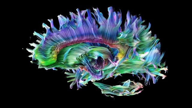
Axons are the minuscule string-like structures that carry information from one place to another. Jones winces as he pulls a hair from his head.
“To put it into perspective, we could stick more than 600 axons inside a single human hair. We’re measuring the movement of water within one of those axons and we’re doing it for everywhere across the whole brain. When you look at it like that, you think, is it possible?
“The scale we’re looking at is way beyond what we could ever actually image. We need to get a thousand times smaller.
"You can get measurements like that, but that means taking the brain out and slicing it, which even highly motivated PhD students aren’t happy to do.”
So they've found another way. By dramatically varying the strength of the magnetic field around the brain, they can detect when a water molecule has moved from one strength of magnetic field to another.
Water molecules move more easily along the fibers than across them. So by detecting the signal from these movements, they can start to see how the axons are structured within the brain, even if the resolution of the scanner isn’t fine enough to see them individually.
“If I have three biscuit tins and I fill one with rice, one with sand and one with pasta shells and then shake them, I can hear that those sand grains are finer than the rice and the pasta shells. I couldn’t exactly tell you about the size of any of them, but I can certainly tell that some are a lot smaller, some are bigger and some are bigger again. And that’s the kind of thing we’re doing.
“We get a collection of signals that are highly influenced by the microstructure. We’re not directly imaging the microstructure itself. We’re measuring its influence on the MRI signal and then using that to build a visualisation.”
Diagnosing mental health
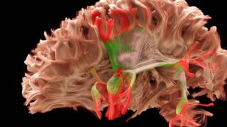
Those properties are thought to play a vital role in a number of conditions. In schizophrenia, it could be that the pattern of connections has gone awry. Multiple sclerosis is thought to attack the myelin, the fatty layer of insulation around the axons, slowing production and causing difficulties with movement. Dementia is associated with a general deterioration of the pathways.
Understanding the causes of these illnesses is a crucial step in improving the lives of the people affected by them. It could mean earlier detection and a more personalised approach to treatment.
Jones sees opportunities to combine improved imaging techniques with emerging technologies like machine learning to recognise patterns in the data.
“If we could combine this with things like genetics, we can identify people at risk and start to stratify people for different treatment regimes.
“You’ve got in vitro diagnostics, which is your bloods, urine and tissue. You’ve got in vivo diagnostics, which is the imaging. And with artificial intelligence and machine learning, you’ve got in silico diagnostics. If you put these all together, you’d have in toto diagnostics, and I think that could be the way to go.”
Does he ever see a scenario where we might screen for mental health problems in the same way we do for some cancers?
“If you can bring the cost down, yeah. I think we’re beyond ten years for that, though, because of the insurance consequences and the psychological downsides.
“I don’t have a crystal ball to say how accurately we’ll be able to predict things in the future, but it’s going to be based on probability, so we’ll be able to say you have an X percent chance of developing a condition.
"If I said to you, you’ve got a 70 percent chance of developing dementia, what will you do with that information? It’s a bit depressing, but what would you do?
“With extremely high likelihood of breast cancer, you have double mastectomies. The options there are obvious. What the choice is for someone who’s been given a high likelihood of a mental health disorder is less clear.”
These are choices we may soon need to make. MRI has come a long way since its development in the 1970s.
“We’re moving away now from saying well, the signal is greater there, or the structure is more organised, or there seems to be less activity. Those are just descriptive words that don’t actually mean very much. We’re starting to look now at the underlying biology.”
The benefits of this approach are already being realised. A common treatment for epilepsy is to go into the brain and chop out a part of the temporal lobe. The problem is that the pathways that connect to the brain's vision centre pass through the same area.
At the Brain Research Imaging Centre, they’re working with a brain surgeon to show where those pathways are in individual patients so that they can be spared in surgery.
It’s these types of collaboration, bringing together experts from different disciplines, that inspired the concept behind the Centre.
Essentially organised around one giant corridor, the diversity of equipment, from big MRI scanners to specialised sleep laboratories, has attracted a diversity of experts.
The Centre's philosophy is to no have no distinct research groups. Instead, people from different disciplines are dispersed throughout the building to maximise the mixing of people and ideas.
“There’s a clinical psychologist who works for the epilepsy team, and I bumped into her in the kitchen. We started talking and now we’ve designed a whole project together where she’s designed a virtual maze for people to walk around, and we’re going to see how the brain responds.”
“It’s just what happens here. The kitchen is an amazing place.”
