Brain scanners
Our brain scanning technology includes structural and functional magnetic resonance imaging, electro- and magneto-encephalography, and transcranial magnetic stimulation.
Magnetic resonance imaging scanner
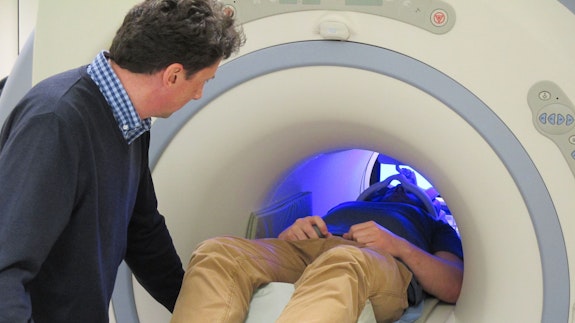
Our magnetic resonance imaging (MRI) scanner provides detailed pictures of the human body.
It uses a very strong magnetic field, generated by a superconducting magnet, together with a radio antenna to "listen" to the electrical signals produced by the hydrogen nuclei of molecules in the body.
The technique is non-invasive and is routinely used in clinical practice.
Magnetoencephalography scanner

Our magnetoencephalography (MEG) scanner can be used to study any aspect of sensory or cognitive function and provides high temporal resolution maps of cortical activity.
It uses sensitive magnetometers to map brain activity, recording magnetic fields produced by electrical currents occurring naturally in the brain.
Transcranial magnetic stimulation facilities
- two dedicated transcranial magnetic stimulation labs
- three Magstim Super Rapid systems for the delivery of magnetic stimulation using a single TMS coil, or multiple coils
- online eye-tracking systems for the monitoring of gaze and eye movements
- a Cambridge Research Systems Visage system for visual stimulus presentation
- a Piezostimulator system for mechanical tactile stimulation
Electroencephalography laboratories

We have two dedicated electroencephalography (EEG) labs, used to study many aspects of sensory and cognitive function, including memory. Each lab contains multi-channel EEG acquisition systems.
We also have facilities for conducting EEG measurements during MRI, used for simultaneous EEG-fMRI.
How they help
Using our MRI scanner, our research found that a technique known as Neurofeedback helps ease the symptoms of depression and Parkinson's disease. Neurofeedback involves patients going into an MRI scanner where their brain activity is continuously measured with functional magnetic resonance imaging (fMRI) and fed back to them.
The Wales Autism Research Centre used our imaging techniques to investigate autism spectrum disorder.
Our equipment has helped discover that some men could be more impulsive, act aggressively, drink and take drugs because they have lower levels of a naturally occurring substance in a specific part of their brain.

Our brain scanning facilities are located in the Cardiff University Brain Research Imaging Centre (CUBRIC).