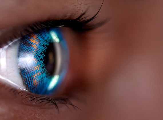
Skip to content
Skip to navigation menu


Three-dimensional MRI scans of avian eyes

Background

Optical coherence tomography (OCT) images of the patient's normal macula and of the retina in the other eye with the macular detachment

Optical coherence tomography (OCT) images of the patient's normal macula and of the retina in the other eye with the macular detachment

Like body size, eye size varies markedly within the animal kingdom. Even within a species, eye size often differs depending on an individual’s age, sex and sometimes, their body size. This variability in eye size is intriguing, given that the component parts of the eye must be perfectly tailored to one another in order for the eye to function as an effective optical system. Mismatching of the eye’s component parts leads to a refractive error: long-sightedness (hyperopia) or short-sightedness (myopia). Currently, little is known about the physiological mechanisms that control the size of the eye and its key optical features, the cornea, lens and vitreous chamber. However, much of the variation is believed to be genetically determined.

  Magnetic resonance imaging can provide non-invasive, high-resolution, three-dimensional information on the structure of biological tissues. MRI is based on the principal that when tissue is placed in a strong magnetic field, the spins of the protons in its hydrogen atoms (mostly in water and fat) go from being randomly oriented to an aligned orientation. This alignment is detected using radio frequency waves, whose modulations are processed by a computer to build up a 3-D representation of the spatial distribution of hydrogen atoms, and thus tissue structure.


MRI movie 1MRI movie 2

We have used a state-of-the-art 9.4 Tesla MRI scanner at Cardiff University to obtain detailed, three-dimensional images of a collection of avian eyes. Chickens from a broiler line (large body and eye size) and a layer line (small body and eye size) were intercrossed for several generations, to provide a set of birds with a wide variety of eye sizes. Blood samples were taken from the birds for genetic analysis, and an MRI scan of one of their eyes was acquired at an “isotropic” resolution of 117 mm. Together, these data are being used to identify genetic variants controlling a wide variety of features relating to avian eye size. These MRI images are now available for use by other researchers interested in avian eye size and shape.

Images here:
http://mri-avian-eyes.cf.ac.uk/ 

Acknowledgements

This work was funded by grants from Cardiff University’s INSRV 'Special Collections' digitisation project and the BBSRC.


MRI movie 3

Further Information and links

The methods used for MRI scanning are described in the following article: Tattersall RJ, et al. (2010) Ex vivo magnetic resonance imaging of crystalline lens dimensions in chicken. Molecular Vision 16:144-153. http://www.molvis.org/molvis/v16/a18/ 


Cardiff University SCHOLAR Special Collections

 http://www.cardiff.ac.uk/insrv/libraries/scolar/index.html