Neuroimaging Huntington's disease
17 May 2017
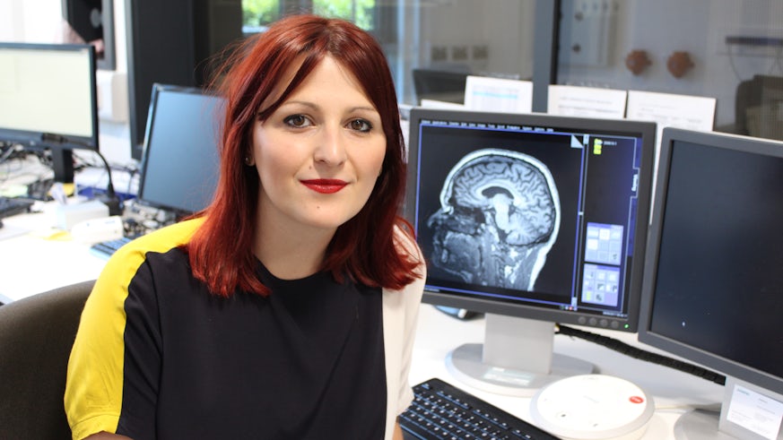
Dr Hannah Furby's work focuses on Huntington's disease, a highly heritable neurodegenerative disease, for which there is currently no cure.
It is characterised by cognitive decline, loss of motor control and psychiatric symptoms, and it affects around 7,000 people in Wales and England.
"My research aims to identify therapeutic interventions that may prevent or reduce the suffering of people diagnosed with Huntington’s disease (HD)" said Hannah, who uses magnetic resonance imaging (MRI) to discover relevant biomarkers of the disease, such as changes in white matter microstrucutre and cerebral blood flow.
"I use these methods to see whether therapeutic interventions, like brain-training, or lifestyle factors, such as physical activity, give rise to subtle changes in the brain that may lead to an improvement in some of the symptoms of the condition."
"Currently, I'm investigating whether completing a home-based computerising brain training programme can bring about disease-related improvements in cognition, and whether this is suggestive of improved biological function."
"There is currently no cure for HD. Whilst some promising drug trials are underway, these still need to undergo further clinical trials in humans. In the meantime, it's important to continue looking for ways to improve the prognosis or delay the progression of the disease, so I am interested in whether home-based interventions like physical activity and brain-training may help."
MRI under-utilised in Huntington's disease research
Compared to other neurodegenerative disorders like Alzheimer’s Disease, there is currently very little MRI research taking place within the field of Huntington’s Disease.
Although it’s prevalence is a lot less (just 1 in 10,000 people), understanding how the toxic effects of the mutant huntingtin gene leads to neurodegeneration can help us to understand the process of neurodegeneration in other diseases.
"Huntington’s disease is unique since we have identified the gene that causes it. This is a dominant gene, which means that if a parent has the disease, their children will have a 50% chance of getting HD themselves." Hannah explains.
Because it is possible to undergo predictive DNA testing, this gives researchers the rare opportunity to assess people earlier in life, prior to symptom onset, when these therapeutics have the greatest chance of improving prognosis. Tracking the same patients over time using techniques such as MRI, can be used to identify subtle alterations in the brain that might take place, even before any behavioural symptoms are observed.
"My research focuses on people with pre-manifest HD. I hope that subtle changes in blood flow or micro-structure that are detectable at this early stage of HD can help us understand other neurodegenerative diseases which are less predictable"
"MRI is a fantastic measure for assessing these longitudinal changes in the brain, but may not always be suitable for HD patients who display a motor symptoms. Head motion can lead to blurring of the scans, making the results much more difficult to interpret. Unfortunately, this is a limitation of MRI research, so that we are often restricted to assessing patients earlier in the disease, or those without head motion."
Changes to brain
Up to 50% of the cell loss expected to occur across the course of the disease happens before any motor symptoms are displayed. The region most affected is the striatum, a component of the basal ganglia, a group of nuclei with a variety of functions, including facilitating voluntary movement.
"Using Diffusion Tensor Imaging, a type of MRI that we specialise in at the Cardiff University Brain Imaging Centre (CUBRIC), we have identified tissues changes in pre-symptomatic HD participants, even in the absence of brain shrinkage in these other regions, suggesting that morphological changes occur before the death of neurons."
The future
Working with neuroimaging specialists at CUBRIC, Huntington's disease researchers are using advanced imaging techniques and developing analysis pipelines to help detect subtle changes in brain physiology.
"Quantifying the physiological effects of cognitive and physical training with MRI will be extremely useful for future research and can be used to guide similar interventions in other diseases" said Hannah.
"I have seen the difficulties that the disease can cause for families and have been inspired by the dedication that patients have when it comes to taking part in research. I hope that the research I do will contribute to this understanding, and in some small way, alleviate the suffering experienced by people affected by the disease."
Get involved
If you're interested in helping with our MRI research into Huntington's disease, contact Hannah:

