Better Understanding of Pancreatic Cancer Will Lead to Earlier Detection for Patients
11 December 2014
Dr Catherine Hogan, Research Fellow at the European Cancer Stem Cell Research Institute, explains why pancreatic cancer is so hard to diagnose and how gaining a better understanding of the disease will result in earlier detection.
Pancreatic cancer is a devastating disease because it has the poorest prognosis of all solid tumours with a 5-year survival rate of less than 5%. Hope for improved patient prognosis lies in early detection and halting the disease at the pre-invasive stages. Early detection can also result in the development of new clinical trials with improved survival. Pancreatic cancer is difficult to diagnose early because it is highly aggressive and we lack suitable screening methods to detect at risk individuals. In recent years our knowledge of the genetics and pathology of pancreatic cancer has improved considerably. However, our knowledge of how this disease begins and develops at the cellular level remains insufficient.
Pancreatic ductal adenocarcinoma (PDAC), the most common form of pancreatic cancer, arises from precursor lesions called pancreatic intraepithelial neoplasia (PanINs). These lesions progress from stage I to stage III and eventually to metastatic PDAC as cells acquire genetic mutations and become transformed. Some of the founder genetic mutations for PanIN formation and progression to PDAC are known. Activating mutations in the oncogene KRas are the first mutation to occur, which transform a normal cell to a pre-cancerous cell. Using tools that map the genetic profile of tumours, researchers have found that human metastatic pancreatic cancers genetically evolve over time. However, late stage tumours remain genetically similar to the initial pre-invasive tumour, suggesting that founder mutations such as oncogenic KRas are required for all stages of disease progression. Using this genetic data researchers have estimated on the timing of development of the various stages of disease, and revealed that on average, a primary tumour expands and grows within the pancreas for a decade before spreading to other organs. This suggests that there is a window of opportunity to detect and diagnose the cancer early, providing we have the right tools. Conversely, a more recent study has revealed that KRas-transformed cells can be detected in the bloodstream and can metastasize to other tissues before a solid tumour has formed. It is possible that although spreading out of the pancreas at very early stages of the disease, these mutant cells once seeded in secondary sites become dormant or grow at a slower rate and are not detected until years later. How these mutant cells are leaving the pancreatic tissue to enter the bloodstream and metastasize to other organs is less clear. More importantly, which cell type is the cell of origin for PanIN formation remains a contentious issue.
At the European Cancer Stem Cell Research Institute, our primary research focus is to gain a better understanding of early pancreatic cancer. In particular, how PanINs initiate and develop following the very first transformation of normal cells, activation of oncogenic KRas. Using a mouse model of pancreatic cancer, work from our collaborator, Professor Owen Sansom at the Beatson Institute of Cancer Research in Glasgow, has shown that the majority of cells expressing mutations in KRas 'disappear' from the pancreas and only a minority of cells stays in the tissue and develop into a tumour.
Using cell culture systems, our research demonstrates that KRas-transformed cells are pushed out of the normal tissue, via a process that requires direct cell-cell interaction with the normal cells. This suggests that the normal cells detect and eliminate the mutant neighbouring cells. Currently, we are exploring whether this is occurring in the pancreas after cells become transformed with mutant KRas.
We are also testing the hypothesis that the mutant cells are escaping the control of the normal cells, and therefore in this context, this process may promote the early spread of the mutant cells. A big question is how do normal cells detect the mutant cells, and currently we are investigating the cell-cell communication signals that may trigger this process.
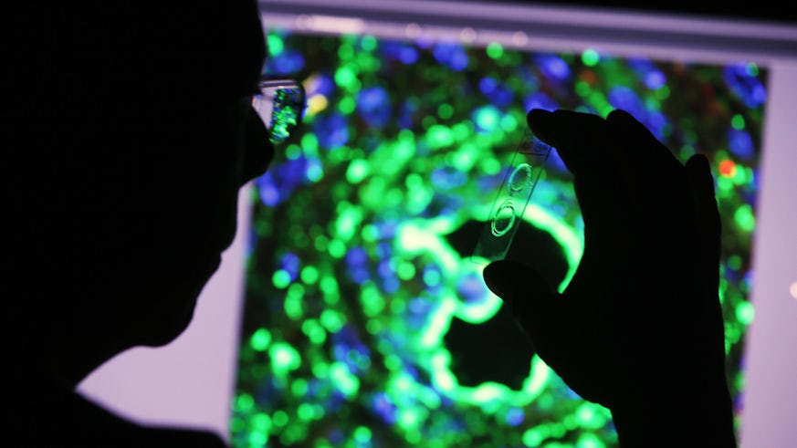
Using fluorescence imaging, microscopy and image analyses software, we aim to examine the relationship between cells within a normal pancreas and compare this with tissue that is precancerous. We can then ask how do different cell types interact, communicate and organise in relation to each other within a developing PanIN. We can also examine whether the physical, structural part of the tissue is changing and ask if this is also playing a role in PanIN progression from stage I to stage III. One of the major risk factors for PDAC is pancreatitis, an inflammatory disease of the pancreas caused primarily by alcohol abuse, which can significantly alter the structure of a tissue. We will address whether and how pancreatitis may alter cell-cell communication between normal and KRas-transformed cells and examine this at the cellular level. This project will be carried out in collaboration with Professor Ole Petersen's team within Cardiff University's School of Biosciences, who are experts in pancreatitis. Together, we hope that our research will add knowledge to our understanding of how pancreatic cancer starts. By understanding the signals that control early disease we may identify new biomarkers and diagnostic tools.