New X-ray Imaging Technique Published in Science by Harris Group
30 Mai 2014
A new X-ray imaging technique developed by scientists from the School of Chemistry at Cardiff University and collaborators from the Diamond Light Source (the UK synchrotron radiation facility) is reported today in the journal Science (DOI: 10.1126/science.1253537).
The research project was led by Professor Kenneth Harris of the Cardiff School of Chemistry, together with Dr Ben Palmer, Greg Edwards-Gau and Dr Benson Kariuki from Cardiff, and Professor Steve Collins and Dr Igor Dolbnya from Diamond Light Source.
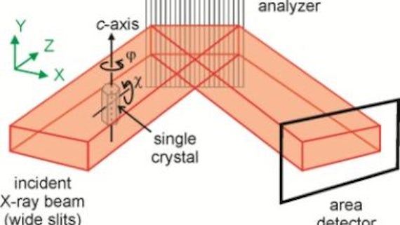
The new technique – called X-ray Birefringence Imaging (XBI) – follows a close analogy in concept to the polarizing optical microscope, which was invented in the 19th Century and is used extensively across the physical, geological and biological sciences. However, rather than polarized visible light, the new technique uses polarized X-rays to interrogate the material of interest, yielding spatially resolved mapping of the X-ray birefringence of the material. X-ray birefringence is sensitive to the orientational properties of individual molecules and/or bonds within a material (thus differing in subtle ways from the optical birefringence that underlies the operation of the polarizing optical microscope).
The Science paper is the first report of an experimental set-up that enables X-ray birefringence measurements to be carried out in a spatially resolved manner, producing a direct image of the X-ray birefringence from all parts of the material under investigation. The application of the X-ray Birefringence Imaging technique is demonstrated by experiments carried out under conditions that selectively detected the orientations of C–Br bonds in brominated organic solids, highlighting the utility and sensitivity of the technique for imaging the local orientational properties of materials, including characterization of changes in molecular orientational ordering associated with solid-state phase transitions. The results also show that X-ray Birefringence Imaging is a powerful tool for identification and characterization of "domain structures" in materials, in which different regions of the material comprise orientationally distinct arrangements of the component molecules, leading to an understanding of the size, spatial distribution and temperature-dependence of the domain structures.
The new technique opens up significant new opportunities for spatially resolved mapping of materials, and is likely to impact on a wide range of scientific fields. Although demonstrated for single-crystal samples, there is no requirement for crystallinity, as X-ray birefringence is sensitive specifically to local molecular orientations. Thus, X-ray Birefringence Imaging could be applied to any material (including liquid or amorphous phases) with an anisotropic distribution of molecular orientations. The technique also has particular utility in cases (e.g. partially ordered materials, multiply twinned crystals, or other materials with complex domain structures) for which the application of diffraction-based techniques is either not straightforward or not feasible.
In addition to providing new insights on the structural anisotropy of a wide range of solid materials, future opportunities for applications of X-ray Birefringence Imaging include studies of liquid crystalline materials and oriented molecules at interfaces. Furthermore, as highlighted in the Science paper, X-ray Birefringence images can be recorded rapidly, leading to very exciting prospects for exploiting the time-resolved capabilities of X-ray Birefringence Imaging. Such applications would allow time-dependent changes in molecular orientational distributions to be probed (for example, studies of the propagation of domain boundaries during phase transitions).