Electroencephalography labs
Watch a video about electroencephalography
We have three dedicated Electroencephalography (EEG) laboratories where we carry out cutting-edge brain research.
How EEG works
Electroencephalography (EEG) is a non-invasive technique used to monitor the electrical activity of the brain. Each individual neuron has an electric potential, but this is far too small to be detected by EEG. Therefore, EEG activity represents the summation of millions of neurons that fire together and are of a similar spatial orientation. The activity we see in the EEG is thought to reflect predominantly pyramidal neurons.
EEG is often performed by applying an elastic cap with electrodes embedded in it to the participant's head. The electrodes are attached to the scalp to record minute electrical activities (down to microvolts). These minimal activities can be amplified, digitised and recorded by a computer for further analysis. EEG is a safe and non-invasive way of measuring brain activity.

Applications of EEG
EEG is useful for both research and clinical applications. It has been widely used to investigate research questions in cognitive neuroscience and psychology. With appropriate experimental designs, the excellent time-resolution of EEG (down to even shorter than 1 millisecond) can be used to investigate various cognitive functions such as: attention, perception, auditory processing, motor preparation, language, and memory.

EEG facilities
We have three EEG labs and one preparation room with dedicated washing facilities. All the labs are equipped with BioSemi ActiveTwo systems, and a passive Brain Vision Quickamp system from Brain Products is also available upon request. All three labs are also equipped with computers which can present stimuli and collect responses from participants via keyboards or response boxes, together with their EEG activity.
The BioSemi ActiveTwo systems are capable of recording up to 128 channels with sampling rate ranges from 2048 to 8192 Hz. The Brain Vision Quickamp system is capable of recording up to 128 channels with sampling rate ranges from 125 to 2048 Hz. Caps for both systems are also available in a range of sizes (52-62 cm). Matlab (Cogent and Psychtoolbox), E-Prime and PsychoPy are available in the labs. Other presentation platforms are available upon request.
EEG recording can also be used simultaneously with MRI, MEG or Brain Stimulation techniques, as well as for sleep studies and developmental studies.
In addition to the EEG systems, we also have a Polhemus Fastrak digitiser system for digitising electrode locations and head shapes. The location information of the electrodes can facilitate studies using source localisations.
Find out more about our EEG equipment.
Apply to use our facilities.
-
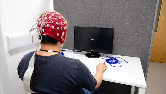
All our EEG labs are equipped with computers that can present stimuli and collect responses from participants via keyboards or response boxes.
-
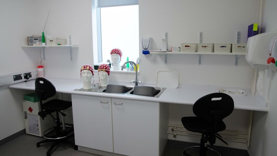
Electrodes are placed on the participant's head in the dedicated EEG preparation room.
-
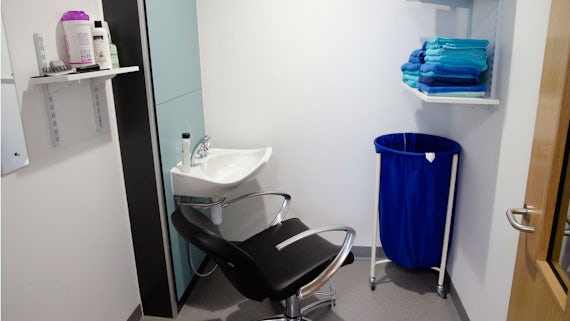
EEG hair washing facilities are used for the removal of electrode gel after testing.
-
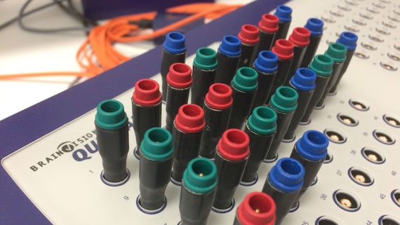
The Brain Vision Quickamp system is capable of recording up to 128 channels.
-
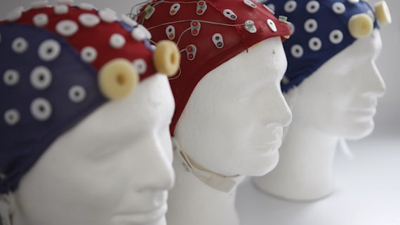
EEG caps
-
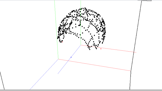
An example of a digitised head shape rendered by the Polhemus Fastrak digitiser.
-
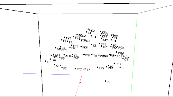
The Polhemus Fastrak digitiser system creates a precise visual representation of electrode locations on the scalp.
Learn more about our research equipment, including the make, model and capabilities, on our database.